Plant Cell vs. Animal Cell: Plant And Animal Cells Coloring Page
Plant and animal cells coloring page – Plant and animal cells, the fundamental building blocks of life, share many similarities, both belonging to the eukaryotic family. However, their distinct roles in the biological world have led to significant structural differences that profoundly impact their functions. Understanding these differences is crucial to appreciating the diversity and complexity of life.
While both cell types contain common organelles such as the nucleus, mitochondria, and ribosomes, key distinctions lie in the presence or absence of certain structures. These differences are not merely cosmetic; they reflect fundamental variations in how these cells obtain energy, maintain their shape, and interact with their environment.
Key Organelle Differences Between Plant and Animal Cells
The following table summarizes the key differences in organelles between plant and animal cells, highlighting their functions and the implications of their presence or absence.
| Organelle | Plant Cell | Animal Cell | Description of Function |
|---|---|---|---|
| Cell Wall | Present | Absent | Provides structural support and protection; maintains cell shape; prevents excessive water uptake. Composed primarily of cellulose. |
| Chloroplasts | Present | Absent | Sites of photosynthesis, where light energy is converted into chemical energy (glucose) using chlorophyll. This process provides the plant with its food. |
| Vacuoles | Present (large central vacuole) | Present (smaller, numerous vacuoles) | Storage compartments for water, nutrients, waste products, and pigments. The large central vacuole in plant cells contributes significantly to turgor pressure, maintaining cell rigidity. |
Functions of Key Organelles
A deeper understanding of the functions of these key organelles further illuminates the differences between plant and animal cells.
The cell wall, a rigid outer layer unique to plant cells, provides crucial structural support, protecting the cell from mechanical damage and maintaining its shape. This is particularly important in plants that lack skeletal systems. The cell wall also regulates water uptake, preventing the cell from bursting due to osmotic pressure. The cellulose composition of the plant cell wall is a defining characteristic.
Exploring the intricacies of plant and animal cells through coloring pages offers a fun, educational experience for kids. For a broader range of animal-themed activities, consider downloading a free resource like this animals coloring book pdf free download , which provides additional engaging options. Returning to the cellular level, remember that the detailed coloring pages of plant and animal cells help children visualize the fundamental differences between these basic units of life.
Chloroplasts are the powerhouses of plant cells, responsible for photosynthesis. Through this process, light energy is captured and used to convert carbon dioxide and water into glucose, a vital energy source for the plant. This ability to produce its own food is a defining characteristic of plants and distinguishes them from animals, which are heterotrophic (relying on external sources for food).
Vacuoles act as storage compartments within both plant and animal cells. However, plant cells possess a large central vacuole that occupies a significant portion of the cell’s volume. This vacuole plays a critical role in maintaining turgor pressure, the pressure exerted by the cell contents against the cell wall. This pressure is essential for maintaining the plant’s rigidity and overall structure.
Animal cells, in contrast, have smaller, more numerous vacuoles that perform similar storage functions but do not contribute significantly to cell rigidity in the same way.
Implications of Structural Differences
The presence of a cell wall, chloroplasts, and a large central vacuole profoundly impacts the overall function and characteristics of plant cells. These structures enable plants to be autotrophic, capable of producing their own food through photosynthesis, and to maintain their structure and rigidity even in the absence of a skeletal system. Animal cells, lacking these structures, are dependent on external sources of energy and rely on different mechanisms for maintaining cell shape and structural integrity.
These fundamental differences reflect the distinct ecological roles and survival strategies of plants and animals.
Coloring Page Design

Designing a coloring page of a plant cell offers a fun and engaging way to learn about the intricate structures within these vital organisms. A well-designed page should not only be visually appealing but also scientifically accurate, reflecting the relative sizes and positions of the various organelles. This accuracy is crucial for effective learning and understanding.
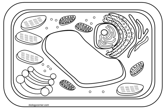
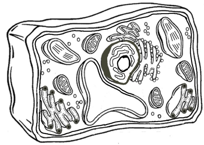
The illustration should depict a typical plant cell, emphasizing its key components. The cell wall, a rigid outer layer providing structure and protection, should be prominently displayed. Within the cell wall, the cell membrane, a selectively permeable barrier, should be clearly visible, slightly smaller than the cell wall. The large central vacuole, a fluid-filled sac responsible for storage and turgor pressure, should occupy a significant portion of the cell’s interior.
The nucleus, containing the genetic material, should be depicted as a relatively large, centrally located structure. Chloroplasts, the sites of photosynthesis, should be numerous and scattered throughout the cytoplasm. The endoplasmic reticulum, a network of membranes involved in protein synthesis and transport, should be shown as a system of interconnected channels. Finally, smaller organelles such as mitochondria (powerhouses of the cell), Golgi apparatus (processing and packaging center), and ribosomes (protein synthesis sites) should be included, although they may be represented at a smaller scale to reflect their relative sizes.
Plant Cell Organelles for Coloring Page
The following list details the organelles that should be included in the plant cell coloring page, ensuring a comprehensive representation of its structure and function. Accurate inclusion of these organelles will provide a more complete understanding of plant cell biology.
- Cell Wall: A thick, rigid outer layer, depicted in a light green or brown.
- Cell Membrane: A thin, flexible membrane just inside the cell wall, shown in a light blue.
- Central Vacuole: A large, fluid-filled sac occupying much of the cell’s interior, colored a pale purple or light blue.
- Nucleus: A large, centrally located structure containing the cell’s DNA, colored a dark pink or red.
- Chloroplasts: Numerous oval-shaped organelles scattered throughout the cytoplasm, colored a bright green.
- Mitochondria: Small, oval-shaped organelles responsible for energy production, colored a dark red or purple.
- Endoplasmic Reticulum: A network of interconnected membranes, shown as a system of interconnected channels in a light yellow or orange.
- Golgi Apparatus: A stack of flattened sacs, represented as small, stacked pancakes in a light brown.
- Ribosomes: Small, dot-like structures found on the endoplasmic reticulum and throughout the cytoplasm, colored a dark grey or black.
Importance of Accurate Organelle Representation
Accurate representation of the size and position of organelles is critical in scientific illustrations because it conveys essential information about the cell’s structure and function. For example, the large size of the central vacuole in plant cells directly reflects its importance in maintaining turgor pressure and storing water and nutrients. Similarly, the numerous chloroplasts emphasize the plant cell’s role in photosynthesis.
Inaccurate depiction can lead to misconceptions about the relative importance and spatial arrangement of organelles, hindering a learner’s comprehension of cellular processes. Illustrations should strive for realism, helping viewers understand the spatial relationships between organelles and appreciate the complexity of the plant cell’s organization. A properly scaled and positioned illustration aids in visual learning and reinforces the understanding of the cell’s structure and function.
Coloring Page Design
Designing a captivating and educational coloring page for an animal cell requires careful consideration of both artistic appeal and scientific accuracy. The goal is to create a visually engaging illustration that accurately represents the cell’s organelles and their spatial relationships, while simultaneously providing a fun and educational experience for the user. The challenge lies in translating the three-dimensional complexity of a cell into a two-dimensional representation that remains clear and understandable.This coloring page will depict a typical animal cell, focusing on its key components.
We’ll use a vibrant color scheme to help distinguish the different organelles and their functions, making the learning process more enjoyable and memorable. The design will aim for a balance between realism and artistic expression, prioritizing clarity and accuracy in representing the organelles.
Animal Cell Organelles for the Coloring Page
The selection of organelles included in the coloring page is crucial for educational effectiveness. Overloading the page with too many organelles can lead to confusion, while omitting essential ones would be misleading. Therefore, a careful selection of the most important and easily recognizable organelles is necessary.
- Cell Membrane: A light blue, wavy line representing the flexible outer boundary of the cell. This will be depicted as a thin, continuous line surrounding all other organelles.
- Nucleus: A large, dark purple circle near the center of the cell, containing a smaller, lighter purple circle representing the nucleolus.
- Cytoplasm: A light yellow background filling the space between the organelles, representing the jelly-like substance within the cell.
- Mitochondria: Several smaller, dark red bean-shaped structures scattered throughout the cytoplasm, illustrating their role as the cell’s powerhouses.
- Ribosomes: Numerous tiny, dark gray dots scattered throughout the cytoplasm, representing the protein synthesis factories.
- Endoplasmic Reticulum (ER): A network of interconnected light green tubes and sacs extending throughout the cytoplasm, showcasing the ER’s role in transport and protein synthesis. The rough ER (with ribosomes attached) could be shown as slightly darker green.
- Golgi Apparatus: A stack of flattened, light orange sacs near the nucleus, illustrating its role in packaging and modifying proteins.
- Lysosomes: Several small, dark green circles scattered in the cytoplasm, representing the cell’s waste disposal units.
Challenges of Representing Three-Dimensional Structure in Two Dimensions
Accurately portraying the three-dimensional structure of organelles within the confines of a two-dimensional coloring page presents a significant challenge. Organelles are not flat, simple shapes; they possess complex three-dimensional forms with intricate internal structures. For example, the endoplasmic reticulum is a vast, interconnected network of membranes, and the mitochondria are bean-shaped with internal cristae. Representing these structures accurately requires creative solutions.
The coloring page will use shading and variations in line thickness to create the illusion of depth and three-dimensionality. The use of color variations within organelles can also help to hint at their complexity. For example, the shading within the mitochondria could indicate the presence of inner folds (cristae). Ultimately, the goal is to convey a sense of the organelle’s three-dimensional nature while maintaining simplicity and clarity for the user.
Educational Applications
Coloring pages, often underestimated, are surprisingly powerful educational tools. They offer a unique blend of creativity and learning, making complex subjects like cell biology accessible and engaging, especially for younger learners. This section will explore the diverse applications of plant and animal cell coloring pages in classroom settings, highlighting their impact on learning and memory.Coloring pages of plant and animal cells provide a hands-on, visual approach to understanding cellular structures.
The act of coloring itself reinforces learning in several ways.
Classroom Applications of Cell Coloring Pages
Coloring pages can be integrated into various classroom activities to enhance understanding of plant and animal cells. For instance, they can be used as a pre-lesson activity to spark interest and activate prior knowledge, or as a post-lesson activity to reinforce concepts and assess comprehension.
- Pre-lesson engagement: Before introducing the topic of plant and animal cells, students can color a blank Artikel of a cell. This allows them to explore the shape and size of a cell, preparing them for more detailed learning.
- Guided learning activity: Students can color a pre-labeled diagram of a cell, matching colors to specific organelles and labeling them as they go. This actively engages them with the material and helps solidify their understanding of each organelle’s function.
- Post-lesson reinforcement: After a lesson on cell structure, students can create their own labeled coloring page, drawing and labeling organelles based on their knowledge. This assessment method provides a visual representation of their understanding.
- Comparative analysis: Students can color both plant and animal cells side-by-side, highlighting the differences and similarities between them. This promotes critical thinking and comparative analysis skills.
- Creative expression: Students can create their own artistic interpretations of plant and animal cells, adding creative elements while maintaining the accuracy of the cell’s structure. This fosters creativity and allows for individual expression.
The Impact of Coloring on Learning and Memory
The act of coloring isn’t merely a passive activity; it actively engages multiple cognitive processes, improving learning and memory retention. The physical act of coloring stimulates different parts of the brain, creating stronger neural pathways associated with the information being learned.
Coloring promotes active recall and reinforces visual learning, leading to improved memory consolidation.
The process of associating specific colors with particular organelles helps students create visual mnemonics, making it easier to recall and distinguish different cellular structures. This multi-sensory approach significantly enhances learning and retention compared to passive learning methods. For example, consistently associating the color green with chloroplasts helps students remember the role of chloroplasts in photosynthesis.
Adapting Coloring Pages for Different Age Groups and Learning Styles
Coloring pages can be easily adapted to suit different age groups and learning styles. Younger students might benefit from simpler diagrams with fewer organelles, while older students can work with more complex diagrams requiring detailed labeling and annotation.
- Younger students (K-2): Simple Artikels of cells with large, easily colored organelles. Focus on basic shapes and colors, avoiding complex labels.
- Intermediate students (3-5): More detailed diagrams with labeled organelles. Introduce simple descriptions of each organelle’s function.
- Older students (6-8): Complex diagrams with multiple organelles and detailed labeling. Include challenges such as matching organelles to their functions or comparing plant and animal cells.
Furthermore, coloring pages can cater to various learning styles. Visual learners benefit directly from the visual representation, while kinesthetic learners engage in the physical act of coloring. Auditory learners can incorporate verbal descriptions of organelles while coloring. Adapting the complexity and activities associated with the coloring pages ensures inclusivity and caters to the diverse needs of the students.
Beyond the Basics

So far, we’ve explored the fundamental structures of plant and animal cells. But the cellular world is far more diverse and fascinating than just these basic blueprints! Many cells specialize to perform specific tasks, developing unique structures to excel in their roles within a larger organism. Let’s delve into the captivating world of specialized cells.
Specialized Cell Types and Their Functions, Plant and animal cells coloring page
Understanding specialized cells helps us appreciate the complexity and efficiency of living organisms. The following table highlights some key examples, showcasing how structure directly relates to function. Imagine the intricate details we could capture in a coloring page dedicated to these unique cellular heroes!
| Cell Type | Organism | Specialized Function | Unique Structural Features |
|---|---|---|---|
| Nerve Cell (Neuron) | Animals (e.g., humans, mammals) | Transmission of electrical signals throughout the body. | Long, slender axons and dendrites extending from the cell body to transmit signals over long distances; numerous synapses for communication with other neurons; myelin sheath (in some neurons) for faster signal transmission. A coloring page could highlight the branching dendrites and the long axon, perhaps even showing the myelin sheath as a spiraling band of color. |
| Root Hair Cell | Plants (e.g., flowering plants, trees) | Absorption of water and minerals from the soil. | Long, thin extensions (root hairs) greatly increase the surface area available for absorption; thin cell walls to facilitate easy passage of water and minerals. A coloring page could emphasize the extensive network of root hairs extending from the main root cell, showing their delicate structure. |
| Guard Cell | Plants (e.g., flowering plants, trees) | Regulation of gas exchange (transpiration) through stomata. | Kidney-bean shape; unevenly thickened cell walls allowing for changes in shape to open and close the stomata; chloroplasts for photosynthesis. A coloring page could depict the guard cells flanking the stoma, showing their change in shape to illustrate the opening and closing mechanism. |
| Red Blood Cell (Erythrocyte) | Animals (e.g., humans, mammals) | Oxygen transport throughout the body. | Biconcave disc shape, maximizing surface area for oxygen uptake and release; lack of nucleus and other organelles to provide more space for hemoglobin. A coloring page could showcase the biconcave shape and perhaps even represent hemoglobin molecules within the cell using a distinct color. |
Adapting Coloring Pages to Illustrate Specialized Cells
Coloring pages can be powerful educational tools. By incorporating accurate representations of specialized cells, we can engage learners in a visual and memorable way. For example, a coloring page featuring a nerve cell could include labels for the axon, dendrites, and myelin sheath, reinforcing their functions. Similarly, a coloring page depicting root hair cells could highlight their extensive network and the process of water absorption.
The use of vibrant colors and clear labeling will make these complex structures more accessible and engaging for children.
