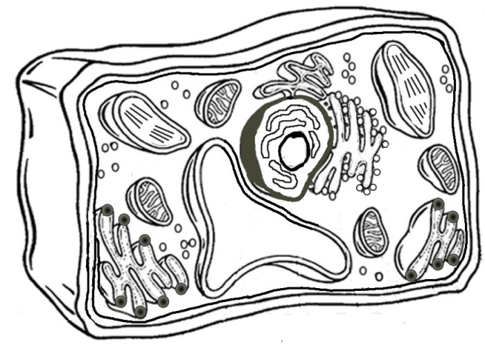
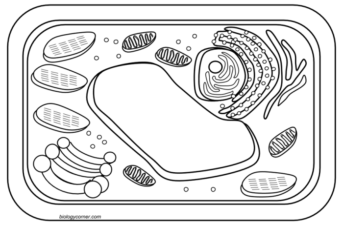
Cellular Structures & Coloring Techniques: Coloring The Cell Animal & Plant

Coloring the cell animal & plant – Right, so we’re diving deep into the nitty-gritty of cell structure and how to get those bad boys looking vibrant under a microscope. We’re talking plant cells versus animal cells – the key differences and the best ways to make ’em pop with colour. Think of it as giving your cells a proper makeover.
The main structural differences between plant and animal cells are crucial when it comes to staining. Plant cells boast a rigid cell wall made of cellulose, a large central vacuole for storage, and chloroplasts for photosynthesis – none of which are found in animal cells. Animal cells, on the other hand, have a more flexible cell membrane and a variety of organelles like lysosomes and centrioles, absent in plant cells.
These differences dictate which stains work best and what structures will be highlighted.
Staining Techniques for Cell Visualization
Various staining techniques are used to visualise specific cell components. The choice of stain depends on the target structure and whether you’re dealing with a plant or animal cell. Different stains bind to different cellular components, producing distinct colours.
| Stain Name | Target Structure | Color Result | Application |
|---|---|---|---|
| Methylene Blue | Nucleus, cytoplasm | Dark blue | Both plant and animal |
| Iodine | Cellulose (cell wall), starch granules | Brown/Dark purple | Plant |
| Eosin | Cytoplasm, other cellular components | Pink/Red | Both plant and animal |
| Crystal Violet | Cell wall (especially in bacteria, but also visible in plant cells), nuclei | Purple | Both plant and animal (though more effective on bacteria and plant cell walls) |
Chemical Processes in Cell Staining
Many staining methods rely on the interaction between the stain and the target structure. For example, methylene blue, a cationic dye, binds to negatively charged components within the cell, such as nucleic acids in the nucleus, resulting in the dark blue staining. Iodine, on the other hand, interacts with starch molecules, forming a complex that appears dark purple or brown.
Eosin, an anionic dye, binds to positively charged components in the cytoplasm, giving a pink or red colour. The chemical nature of the stain and its interaction with the cellular components is key to successful staining.
Comparison of Staining Methods
The effectiveness of different staining methods varies depending on the target organelle. For example, iodine is incredibly effective at highlighting the cell wall and starch granules in plant cells, while methylene blue provides a general overview of the nucleus and cytoplasm in both plant and animal cells. Eosin is useful for differentiating cytoplasmic structures, but may not highlight specific organelles as effectively as other stains.
The choice of stain often involves a bit of trial and error, depending on the specific research question and the desired level of detail. For instance, if you’re focused on visualizing the intricate structure of the plant cell wall, iodine is your go-to; if you want a quick overview of the cell’s basic components, methylene blue is a solid choice.
Microscopic Observation & Image Creation

Yo, peeps! So you’ve got your cells all stained up and ready to go – now it’s time to get those bad boys under the microscope and snap some seriously sick pics. This ain’t no amateur hour; we’re talking pro-level cell imaging. Let’s get into the nitty-gritty.
Getting a good look at these tiny dudes requires a bit of skill and the right equipment. We’ll cover preparing your slides, getting the best shots, and dealing with those pesky transparent structures that love to hide. Think of this as your ultimate guide to cell-snapping mastery.
Preparing a Plant Cell Slide for Microscopic Observation
Right, let’s start with the plant cells. First things first, you’ll need a fresh sample – think a bit of onion skin or a leaf cross-section. Gently place your sample onto a clean microscope slide, adding a drop of stain (like iodine or methylene blue) to highlight those cellular structures. Then, carefully lower a coverslip onto the sample, avoiding air bubbles.
Too many bubbles? Gently tap the coverslip to release them. Boom! You’re ready to rock.
Preparing an Animal Cell Slide for Microscopic Observation
Animal cells? Similar vibe, but different source material. You could use a cheek swab (yup, scrape the inside of your cheek!), or blood, or even a prepared slide from a lab supply. Spread your sample thinly on the slide. Add your stain –methylene blue is a popular choice here– and gently place a coverslip on top.
Again, avoid those pesky air bubbles. This prep work is crucial for a clear view.
Capturing High-Quality Images of Stained Plant and Animal Cells, Coloring the cell animal & plant
Now for the fun part: snapping those killer cell shots. Optimal lighting is key – even, bright light will minimise shadows and enhance contrast. Start with lower magnification to get an overview of the sample, then crank it up for those detailed close-ups. Use your microscope’s settings to adjust brightness, contrast, and sharpness. Take multiple images at different magnifications, ensuring each image is in sharp focus.
Remember, patience is a virtue here – take your time and experiment with the settings.
Overcoming Challenges in Imaging Transparent Cell Structures
Transparent cells? Total nightmare for imaging, right? But don’t sweat it. There are techniques to make those sneaky structures visible. Phase-contrast microscopy is a game-changer – it enhances contrast by manipulating the light waves passing through the sample, making those transparent parts pop.
Another technique is differential interference contrast (DIC) microscopy, which uses polarised light to create a 3D-like image with even better contrast. These methods essentially trick the light to make the invisible visible.
Learning about cell structures? Coloring animal and plant cells can be a fun and educational activity for all ages. But if you’re looking for something a bit different after mastering those diagrams, you might be interested in exploring other creative coloring options, like the more mature themes found on sites such as coloring pages suggestive anime. However, remember to always choose coloring activities appropriate for your age and interests.
Returning to the educational side, detailed cell coloring helps reinforce understanding of organelles and their functions.
